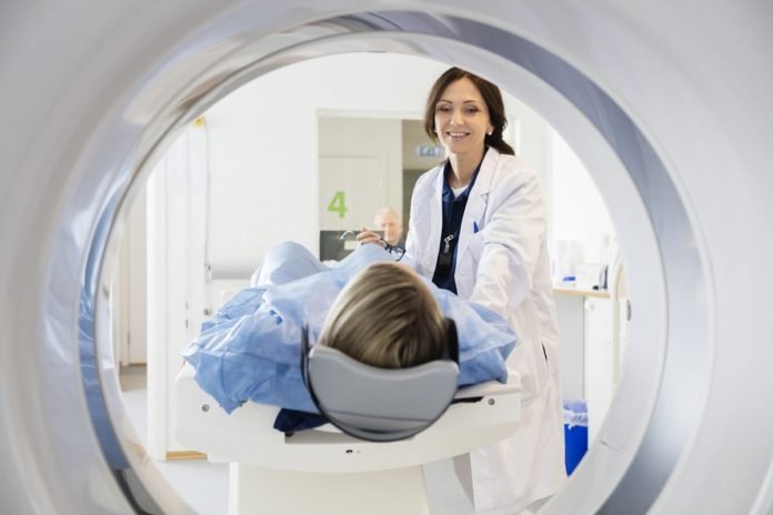
Presently, there are numerous ways mankind can see beyond what the naked eye can, thanks to technology. Imaging technology has been an extremely useful work of technology for the last decades. While image scanners are typically encountered in airports to maintain everyone’s safety and security, imaging technology is even more common in the medical industry.
Whenever an individual notices symptoms of a serious disease, particularly cancer, medical professionals initially recommend having a full body scanning/screening using AI. But, what do we know about full body scan technology, and how has it improved over the years?
How Full Body Scans Save Lives
How does a simple X-ray, magnetic resonance imaging (MRI), or computed tomography (CT) scan image lead to disease treatment? Essentially, medical imaging is an advantageous medical tool that saves many people’s lives, especially of patients who are in the early stages of their diseases. Medical imaging lays the groundwork for physicians to assess what’s going on inside the body, which can’t be assessed by basic medical examinations like vital sign checks and visual examinations.
Because doctors have a clearer idea of the abnormalities occurring in the body, they can come up with a treatment plan that is relatively cheaper, more effective, and more efficient. In case there’s an ongoing treatment, physicians can develop a better and more efficient treatment plan based on the medical imaging findings.
Most importantly, people with higher risks for diseases can take full body scanning on a regular basis as a preventative method. In this way, medical professionals can detect an illness as early as possible, increasing the possibilities of a successful and less invasive treatment with lower medical costs.
Full Body Medical Imaging Through The Years
Medical imaging has been on a long journey of medical development and innovation. Presently, the medical industry can easily access and utilize various medical imaging modalities such as MRI, X-ray, CT scan, positron emission tomography (PET) scan, and ultrasound, and be able to use them for accurate diagnosis.
To understand how medical imaging was developed, take a trip down memory lane and explore body scanning through the years:
First X-Ray By Röntgen in 1895
While conducting an experiment for his research, German mechanical engineer Wilhelm Röntgen accidentally discovered that it’s possible to look through the skin of his hand. He was operating the cathode ray generator, and he saw the bones in his hands on a photographic plate. To confirm his discovery, Röntgen also experimented with his wife’s hand, successfully imaging her bones along with her wedding ring.
Surprisingly, Röntgen simply named it “X-ray” as he wasn’t certain what it was made of. Not long after, European and American laboratories replicated his work with a 3-hour radiograph that involved imaging of objects instead of the human body, such as rings, pins, and opaque metal bullets.

Thanks to the x-ray discovery, physicians then started exploring the medical applications of radiographs, and these are what they found:
- Improved medical assessment of skeletal trauma
- Vital for military medicine in WW1
- Served as treatment for gas gangrene
- Detection of tuberculosis and lung lesions
- Diagnosis of abnormalities in the gastrointestinal tract
The Ultrasound
Although x-rays were a medical milestone, researchers found out that the radiation emitted by x-ray procedures posed long-term adverse effects. But, many people still pursued using this technology due to fascination, even without medical prescription. For instance, obstetricians utilized ultrasound for monitoring a fetus during pregnancy, unmindful of the adverse effects
When the 1930s came, radiation safety procedures were implemented, and the use of radiation technology was more regulated for medical use. Radiation safety was particularly for when imaging infants and children as their premature tissues are more susceptible to radiation-induced damage.
The biggest advantage of utilizing ultrasound versus x-ray is the absence of ionizing radiation in exchange of sound waves. Also, ultrasound machines are portable, allowing physicians to use them by the bedside.
Aside from obstetric purposes, ultrasound also opened the possibilities in different industries:
- Studies on echocardiography encompassing cardiac structures and functions
- Treatment of neurological disorders
- Detection of cracks in metal structures
- Breaking of stones in the kidney called lithotripsy
- Accelerated bone fracture healing using low-intensity pulsed ultrasound (LIPUS)
MRI Discovery by Damadian in 1970s
Before being labelled as magnetic resonance imaging (MRI), this imaging modality was first explored as nuclear magnetic resonance (NMR) spectroscopy in 1945. In NMR spectroscopy, a secondary oscillating magnetic field creates a resonance of an atom’s nucleus, which aids in observing physical, chemical, and biological properties of matter.
Then, in 1969, American physician Raymond Damadian found out that cancerous and non-cancerous tissues can be differentiated through the speed in which the signals were returned, with the cancerous tissues taking much longer. With the help of magnetic resonance, he was also able to determine cancerous cells as they carry more hydrogen atoms in them. He designed the first whole-body MRI scanner called “The Indomitable” in 1977.
Today, the medical applications of MRI are unbeatable. This procedure is non-invasive, and continues to widen its scope and use:
- Diseases of the abdominal organs
- Evaluation of endometriosis, fibroids, and conditions involving the pelvis in women
- Joint abnormalities or injuries in the knee and back
- Brain and spinal cord abnormalities
- Screening of breast cancer in men and women
CT Scan by Hounsfield in the 1970s
The last full body scanning modality discovered in the list is the popular computer tomography (CT) scan. The first creator of the CT scanning technology, English electrical engineer Godfrey Hounsfield developed the idea while he was on vacation. According to him, you can see through one object by taking its x-rays separately in varying angles. These images will look like “slices” that can be assembled together to make an image of a body part.
The first medical application of CT scan occurred in 1971 to a patient with a brain tumor. As it wasn’t too long after the machine was developed, it took them a few days to reconstruct the image of the head.
On another end, CT scan results in this present time can be obtained almost immediately, and it boasts the following medical applications:
- Observation of health conditions like heart disease, cancer, liver masses, and emphysema
- Assists in locating infection, blood clot, tumor, and excess fluid
- Utilized in treatment procedures and plans, including radiation therapy, surgery, and biopsy
- Detect bleeding and internal injuries caused by vehicular accidents
The Future of Radiology: Final Thoughts
Radiology and its various modalities are a remarkable technological contribution to the medical world, and it undoubtedly has a bright future ahead. Through years of medical imaging utilization and development, the medical industry works on incorporating artificial intelligence (AI) to come up with precise medicine and treatment plans for patients, equipped with improved diagnostic capacity and efficiency. Molecular imaging and genomics and enhanced interventional radiology are also on the way.


My body always in pain. haw can solve this problem?
regard
Comments are closed.